For clinicians

Aortic hemodynamics from anatomical imaging.

Clinical goal

More personalized care through aortic hemodynamics.

Anatomical context
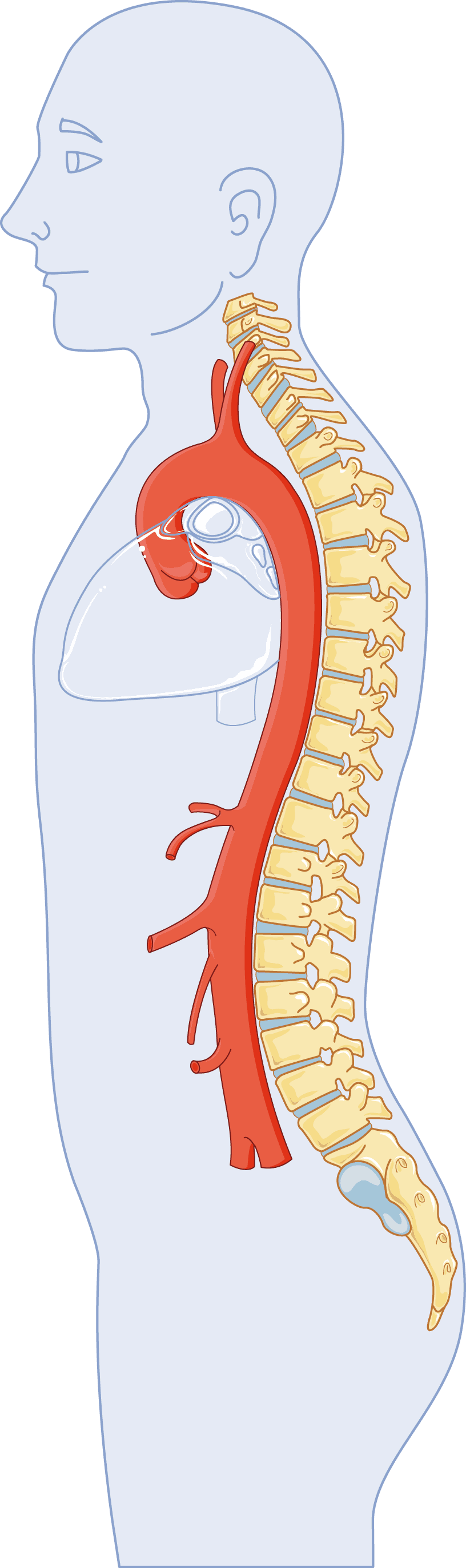
Thoracic aorta

The portion of the aorta located in the chest.

Image adapted from Servier Medical Art, licensed under CC BY 4.0.
Imaging pathway
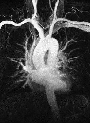
MRI
Company-supplied MR angiography example showing the thoracic aortaMRA image
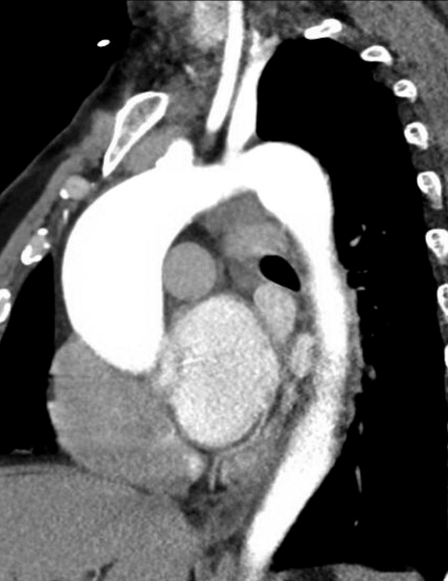
CT
Company-supplied CT angiography example showing the thoracic aortaCTA image
Aortic hemodynamicsPatient-specific blood-flow information

Clinical foundation

The factors clinicians already use

Aortic size and growth remain central to assessment. Clinicians also consider symptoms, family history, genetic findings, valve anatomy, body size, disease phenotype, and other patient-specific factors.

Development focus

Hemodynamic information from anatomical imaging

Third Coast Dynamics is developing technology to derive patient-specific three-dimensional velocity information and selected hemodynamic measures from anatomical CT or MR images. Research is ongoing to better understand how these factors can be incorporated for more individualized aortic risk assessment and management strategies.

Review the peer-reviewed evidence

Connect with us

Connect with our clinical team.

We are dedicated to working alongside radiologists and cardiologists to evaluate how advanced aortic hemodynamic data may support future personalized care strategies.

join the conversation